|
|
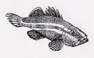
CENTENARIAN SPECIES AND
ROCKFISH PILOT STUDIES
continued
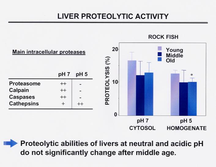
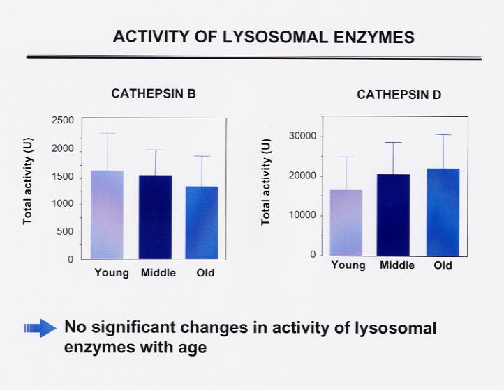
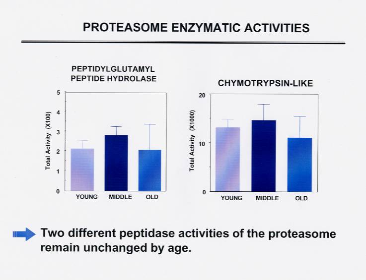
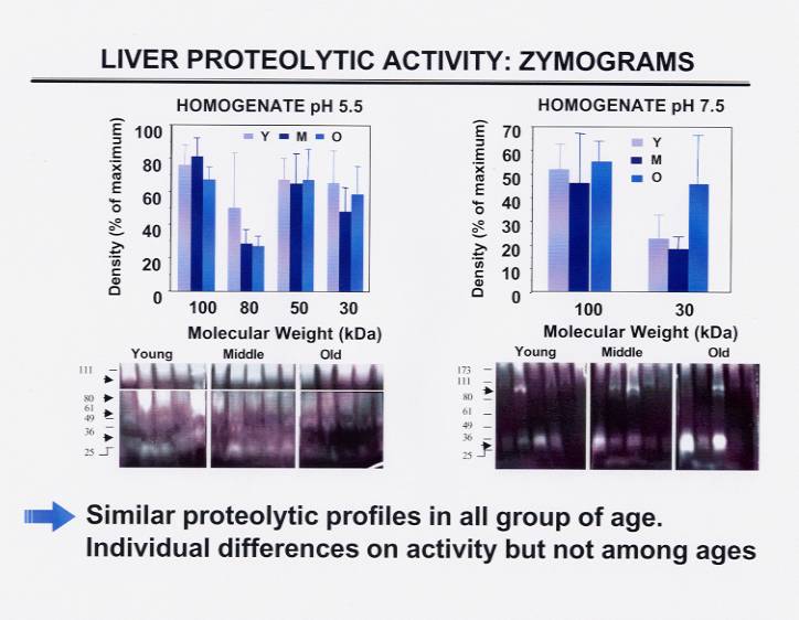
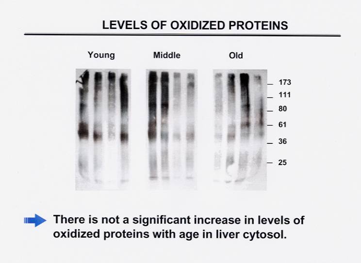
(9 - October 2000) Lysosomal markers of intracellular proteolytic activity. Ana Maria Cuervo, Albert
Einstein College of Medicine, Bronx, NY. In a very extensive pilot study, Ana Maria analyzed levels and activity of
several lysosomal components in liver of different aged rockfish. She sought to determine
if increased protein turnover might contribute to the preservation of systems for removal
of damaged proteins and consequently to better cellular functioning (see Cuervo 2000 for
protocol). In addition she compared levels of selected substrate proteins, and ran
zymograms to analyze specific protease activities (shown are figures for lysosomal
enzymes, proteasome enzymatic activities, zymograms and oxidized proteins). Her age groups
of five samples each were young (14-23 years old), middle (27-37 years old), and oldest
(43-77 years old).
She did not find significant differences in the activity of proteases between age groups,
suggesting that the normal levels of protein degradation found in the oldest animals
results directly from the normal functioning of their proteolytic systems. A standardized
method to detect the presence of oxidized amino acid residues in proteins also showed no
increase with age. She concluded that the groups analyzed here did not exhibit the
dramatic decline in protein degradation with age described in other species such as
rodents. 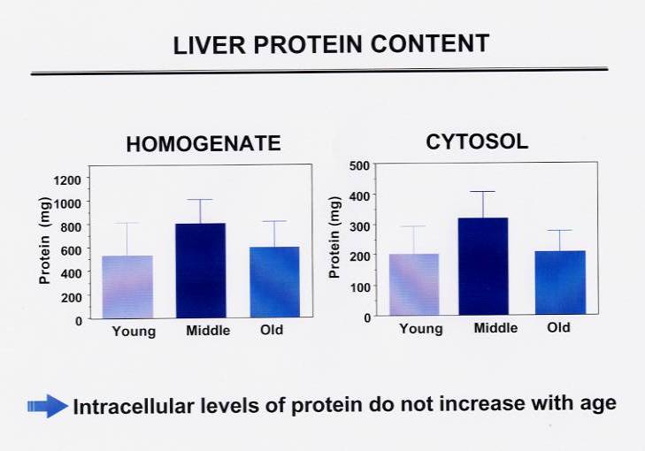
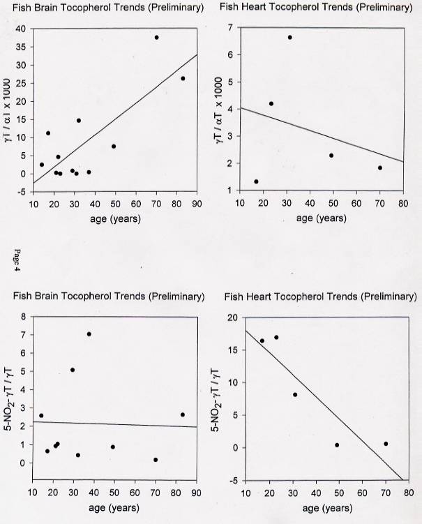
(10 - September 2000) Tocopherol derivatives. Robert A. Floyd, Free Radical Biology and Aging Research
Program, Oklahoma Medical Research Foundation, Oklahoma City, OK. Bob analyzed tocopherol
derivatives in rockfish brain and heart up to 83 years old (Hensley et al 2000 for
protocol). He found unexplained differences in two comparisons, albeit with significant
scatter in the data points (shown are tocopherol comparisons). In gamma over alpha
tocopherol and 5-nitro gamma over total gamma measurements, brain tissue showed a positive
correlation with age or no trend, respectively, whereas heart tissue showed an inverse
relationship with both.
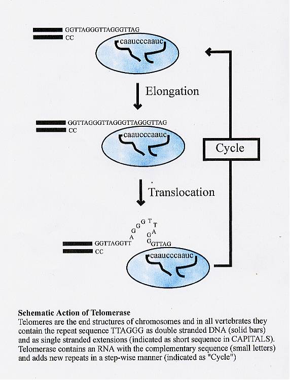
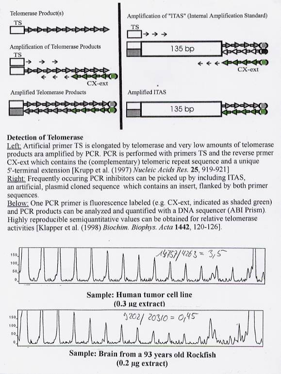
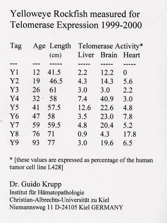
(11 - September 1999) Telomerase expression . Guido Krupp, Institute for
Hematopathology, Christian-Albrechts University, Kiel, Germany. Guido and Wolfram Klapper analyzed
telomerase expression in brain, heart and liver tissues from rockfish up to 93 years old.
According to the telomere hypothesis, DNA replication leads to telomere shortening,
resulting in a cellular mitotic clock (Klapper, Krupp et al 1998a, 1998b). Telomerase
resets it by telomere synthesis. Since most rockfish grow throughout their life, they must
perform continuous cell proliferation. For maintaining this cell proliferation capacity,
telomerase should be active in cells of all somatic tissues, irrespective of fish age.
Results of the pilot study confirmed this expectation: in all three tissues, significant
telomerase activity was detected (shown are figures depicting the telomerase cycle, and
chart comparing expression in rockfish brain, heart and liver tissues). Most importantly,
there was no age-dependent change in expression. Unanswered questions of interest are the
length of rockfish telomeres, the correlation of telomerase activity with cellular
proliferation, and quantification of levels of apoptosis.
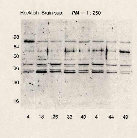
(12 - May 1999) Anion exchange protein in relation to Alzheimer's disease.
Giel Bosman, Department of
Biochemistry, Faculty of Medicine, University of Nijmegen, The Netherlands. Giel
researched anion exchange (AE) proteins in rockfish brain and heart over 90 years old
(Bosman 1997 for protocol). These proteins are known in humans to increase with aging, and
especially with degeneration in Alzheimer's disease-affected brain areas. Elucidation of
the molecular nature of these changes, and the underlying mechanisms, can lead to insight
into the processes that govern aging- and degeneration-associated perturbation of membrane
integrity. Immunoblots showed several reactive protein bands that had approximately the
same molecular weights as those observed in human brain tissue (shown is immunoblot; the
vertical axis is the apparent molecular weight of marker proteins, the horizontal axis is
the rockfish tag numbers). In a preliminary analysis, there appeared to be a decrease in
total AE protein expression with age, in at least one out of three protein bands in both
rockfish brain and heart tissue. A larger sample size will be needed to clarify if this is
an age-dependent effect.
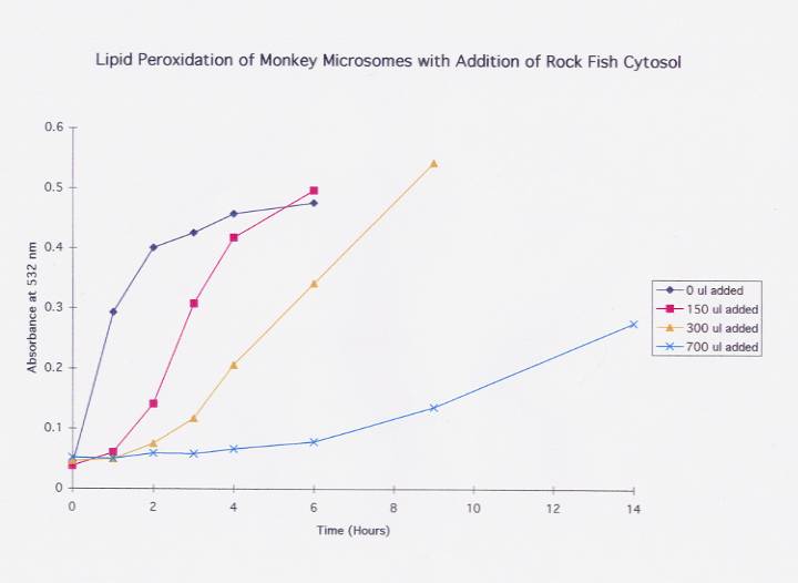
(13 - May 1997) Oxidative damage. David
E. Williams, Linus Pauling Institute, Oregon State
University, Corvallis, OR. David exposed both Rougheye and Yelloweye rockfish liver
samples up to 101 years old to oxidative damage (Kelly et al 1992 for protocol). He found
the generation of TBARS (a marker of lipid peroxidation) was dramatically reduced compared
to rat or monkey liver microsomes (this experiment is shown below, followed by a figure
showing dose-dependent effect of rockfish cytosol protection). Paradoxically, the
polyunsaturated fatty acid (PUFA) content (and hence intrinsic oxidizability) of rockfish
is relatively high compared to trout, for example, suggesting that rockfish have
additional protection from oxidative reactions. Attempts to duplicate this research a year
later with samples that had been stored at -80 degrees C. were unsuccessful. Dr. Williams
suspects the protection may be thermolabile, possibly a protein or peptide.

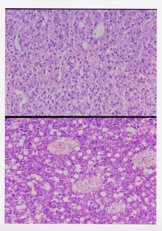
(14 - May 1997) Histology.
Jerry D.
Hendricks, Department of Food Science and Technology,
Oregon State University, Corvallis, OR. Jerry performed histological examinations of
young-, medium- and older-aged rockfish spleen, liver and kidney. He found an increase of
melanomacrophage centers in older specimens, although the physiological consequences of
these melanomacrophage centers does not seem to affect survival (shown in figure is liver
from a 15 year old rockfish in top frame, and an 83 year old rockfish in bottom frame,
both 25X). No other cellular indicator existed to differentiate between the cells of young
and old rockfish, which ranged over 80 years between youngest and oldest samples.
 |